echo|case is an echocardiographic "showcase", intended to present pregnant, visual
impressive echocardiographic findings with high didactical value, seen from the point of view and experience of different
echocardiographists.
This is not a section for case reports, namely, it is not destined for unusual echocardiographic cases or cases of
excepcional occurrence.
Despite of the tremendous advances of echocardiography, auscultation still plays a major role in cardiac examination.
One appropriate indication of echocardiographic examination is: "Initial evaluation of murmur in patients for
whom there is a reasonable suspicion of valvular or structural heart disease." [1]
Images that follow correspond to a 70 year-old patient was appointed to echocardiographic examination for having a
loud systolic murmur, with suspicion of aortic stenosis. The examination revealed a mild to moderate thickened
tricuspid aortic valve with normal opening and velocities, but with evidence of cusps vibrations in the B-, M-mode and
Doppler examination. Innocent murmurs are murmurs without clinical relevance. Their prevalence may be almost as high as 50 % of all
examinations. [2]
Acoustic Doppler artifacts are superimposed Doppler signals, maybe due to overtones produced by
structural vibrations and are not rare in case of anatomically altered heart valves. They can be auscultated as loud,
sometimes as harsh murmurs, sometimes as musical accessory sounds with the stethoscope and they can also be
registered with spectral and acoustic Doppler ultrasound. [3]
Left:
aortic valve from the apical three-chamber view. Aortic valve cusps are moderately
thickened, but show a normal opening move- ment in systole.
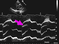
Right:
M-mode display of aortic valve cusps, as seen from the parasternal long axis view. Notice
the fine and regular vibrations of leaflet tips (arrow).
Left:
M-mode moving display of the non-coronary aortic cusp, showing vibrations, as seen from the apical
three-chamber view.
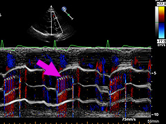
Right:
these cusps vibrations produce also a flow turbulence during the systole, as seen here with
the color M-mode display.
Left:
flow turbulence (arrow) in the color M-mode display of the non-coronary aortic cusp.
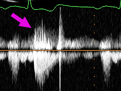
Right:
also Doppler artifacts can be seen (arrow) in the spectral CW Doppler display. This patient
had normal opening of the aortic valve in B- and M-mode, as well as normal systolic velocities (1.6 m/s).
References
1. Douglas PS et al. ACCF/ASE/ACEP/ASNC/SCAI/SCCT/SCMR 2007 appropriateness criteria for transthoracic
and
transesophageal echocardiography. J Am Coll Cardiol 2007;50:187-204.
2. Movahed MR, Ebrahimi R. The prevalence of valvular abnormalities in patients who were referred for
echocardiographic examination with a primary diagnosis of "heart murmur". Echocardiography 2007;24:447-451.

 ORC
ORC