
|
||||||
|---|---|---|---|---|---|---|
| Echocardiography 5 minutes before starting
|
||||||
Examples of pathological findings |
||||||
|
—Echocardiographic examinations |
—Cardiac function and PA pressure |
—Examples of pathological |
||||
|
Aortic dissection
Guidelines and Standards Multimodality Imaging of Diseases of the Thoracic Aorta in Adults, 2015. |
||||||
|
||||||
|
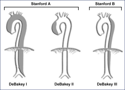
Standford A |
||||||
Left:
dissection membrane starts approx. 2 cm above the aortic valve, the ascending aorta is aneurysmatic.
|
||||||
Left:
cross-section of the ascen- dant aorta in the same case. Notice the intramural hematoma previous to start
of dissection membrane.
|
||||||
|
Standford B |
||||||
Left:
descending aorta short after intersection from the aortic arch. Flat, wall-adherent plaques.
|
||||||
Left:
more distal, a dissection membrane with entrance tear can be seen. False lumen at the bottom of the image.
|
||||||
Left:
dissection membrane with entrance tear and PW-Doppler flow display.
|
||||||
©
Derliz Mereles |
||||||
|
echobasics | free echocardiography tutorial online since 2004 |
||||||
 ORC
ORC