echo|case is an echocardiographic "showcase", intended to present pregnant, visual
impressive echocardiographic findings with high didactical value, seen from the point of view and experience of different
echocardiographists.
This is not a section for case reports, namely, it is not destined for unusual echocardiographic cases or cases of
excepcional occurrence.
Identification of left ventricular thrombus by contrast echocardiography
Prof. Dr. Alexander Hansen, MD
Medizinische Klinik II
Kardiologie, Angiologie, Pneumologie
Klinik Kösching
Krankenhausstr. 19
85092 Kösching
Telefon +49 (08456) 71-431
Telefax +49 (08456) 71-422
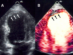
A 64-year-old man with history of coronary artery disease and anterior myocardial infarction was referred
to the echocardiography laboratory for routine evaluation of left ventricular performance. Transthoracic
two-dimensional echocardiography demonstrated a moderate impaired left ventricular function with apical
akinesia. A flat echodense structure was visualized in the apex (arrow) which was suspicious for the
presence of a thrombus (Fig A).
Power Doppler imaging was performed after bolus injection of 0.5 mL a commercially available contrast agent (Fig B). A marked
"perfusion defect" confirmed the diagnosis of an apical thrombus. Therefore, the use of
myocardial contrast imaging improves the diagnostic power of echocardiography for identification of
thrombus.
Left:
Echodense structure on transthoracal echocardiography in the apex of left ventricle (arrows).
Myocardial contrast echocardiography shows an apical thrombus without opacification.
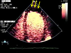
Right:
ECG triggered myocardial contrast echocardiography (MCE) examination shows right ventri- cular,
left ventricular volumes and myocardial phases.
Myocardial Contrast Echocardiography:
MCE is based on the use of echocardiographic contrast agents with inert gas microbubbles, that are small and stable
enough to cross pulmonary circulation and flowing into the left atrial and ventricular cavities, finally enhancing
sonographic view of the coronary circulation.
Intravenous injection of commercially available contrast agents is safe. Microbubbles remain entirely within the
vascular space. Interaction between ultrasound and microbubbles
produces energy with potential effects on tissue for inertial cavitation and acoustic current production.
These effects are interesting for the therapeutic applications of contrast echocardiography, but they do not appear
to have clinically relevant effects.
Contrast echocardiography is considered to be an extension of the standard echocardiographic examination, but requires
additional training, as well as technological upgrading, in order to obtain satisfactory results.
Left:
After injection, the right ventricle is first opacified. After crossing the lung, the
left atrium and ventricle, and finally the coronary circulation at the myo- cardial phase.
Right:
A high energy ultrasound flash destroy the microbubbles, allowing afterwards the rapid filling ot the
coronary bed with new ones. The region with low or absent myocardial perfusion can be detectec
is this way (arrows).
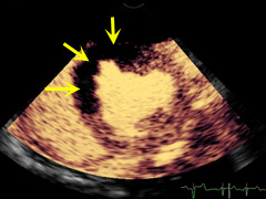
Left:
Experimental case showing an extense myocardial region with absend opacification after ligature of a coronary
vessel.
Right:
As denoted here in this image (arrows).
References
1. Hansen A, Kumar A, Wolf D et al. Evaluation of cardioprotective effects of recombinant soluble P-selectin
glycoprotein
ligand-Ig (rPSGL-Ig) in myocardial ischemia-reperfusion injury by real-time myocardial contrast
echocardiography. Journal of the American College of Cardiology 2004;44:887-891.
2. Hansen A, Johansson BL, Wahren J et al. C-peptide exerts beneficial effects on myocardial blood flow and function
patients with type 1 diabetes. Diabetes 2002;51:3077-3082.
3. Wolf D, Reinhard A, Krause U et al. Stem cell therapy improves myocardial perfusion and cardiac synchronicity:
new application for echocardiography. Journal of the American Society of Echocardiography 2007;20:512-520.
4. Bibra von H, Bone D, Niklasson U et al. Myocardial contrast echocardiography yields best accuracy using
quantitative
analysis of digital data from pulse inversion technique - comparison with second harmonic imaging
and harmonic
power Doppler during simultaneous dipyridamole stress SPECT studies.
Eur J of Echocardiography 2002:3:271-282.

 ORC
ORC