
|
||||||
|---|---|---|---|---|---|---|
| Echocardiography 5 minutes before starting
|
||||||
Echocardiographic examinations |
||||||
|
—Echocardiographic examinations |
—Cardiac function and PA pressure |
—Examples of pathological |
||||
| Strain Imaging: assessment of myocardial mechanics | ||||||
|
Guidelines and Standards Evaluation of Cardiac Mechanics Consensus Statement, 2019
|
||||||
|
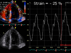
Normal strain: values of myo- cardial deformation at systole lie here around -25 % on the lateral segments of the left ventricle. Color encoded dynamic bidimen- sional image helps to visualize strain, red stands here for -20 %. |
|||||
|
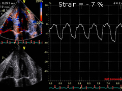
Pathological strain: values of myocardial deformation at systole lie here around -7 %, being consequently very reduced. This case is a proven myocardial invol- vement in systemic amyloidosis. |
|||||
|
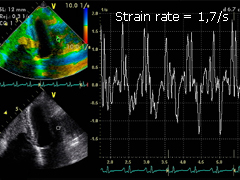
Normal strain rate: values of diastolic myocardial deformation in this normal heart lie between 1.3 and 1.7/s. |
|||||
|
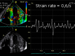
Pathological strain rate: this former amyloidosis case shows also a severe impairment of longitudinal diastolic myocardial deformation, with values around 0.6/s. |
|||||
|
2D Speckle-Tracking Strain The longitudinnal strain in this case is normal. In this case, the longitudinal strain is mildly reduced, especially septal basal. Here is an example of a moderate reduction of the longitudinal strain, especially mediobasal, in a case of cardiac compromise by systemic light-chain amyloidosis. The apical segments show normal values (relative apical sparing). The longitudinal strain is in this case severely diminished, especially mediobasal, in a case of cardiac compromise by systemic light-chain amyloidosis. The longitudinal strain in this case is reduced, especially medioapical, in an apical form of hypertrophic cardiomyopathy. Alternative methods for the assessment of longitudinal strain Feature-Traking Strain is comparable to 2D Spekle-Tracking Strain: Left ventricular mechanics assessed by two-dimensional echocardiography and cardiac magnetic resonance imaging: comparison of high-resolution speckle tracking and feature tracking, 2016 The longitudinal Unidimensional Strain (ULS) is comparable to 2D Speckle-Tracking Strain: Unidimensional Longitudinal Strain: A Simple Approach for the Assessment of Longitudinal Myocardial Deformation by Echocardiography, 2018 Longitudinal LV function may be assessed by the measurement of the anterior movement of the aortic root in systolen: Pathophysiological background and prognostic implication of systolic aortic root motion in non-ischemic dilated cardiomyopathy, 2019 |
||||||
©
Derliz Mereles |
||||||
|
echobasics | free echocardiography tutorial online since 2004 |
||||||
 ORC
ORC